Version 2.0
Imagine having the ability to view internal organs in 3D that are
comprehensive patient-specific. BioMedX is making this a reality
with Version 2.0 of the X³ 3D Physics Engine through Data Merge.
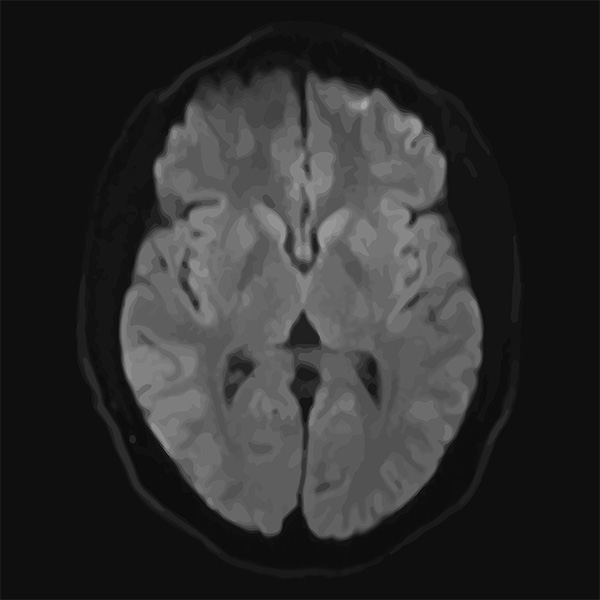
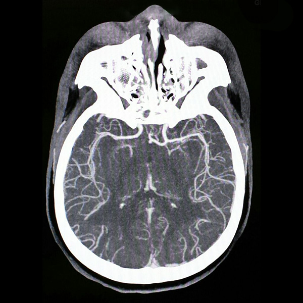
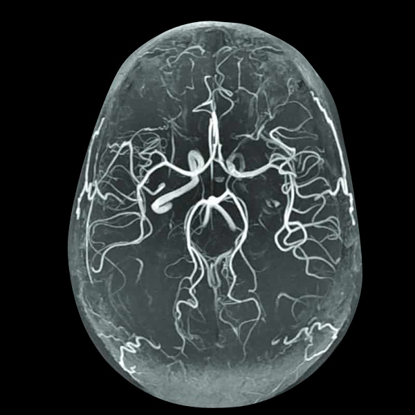
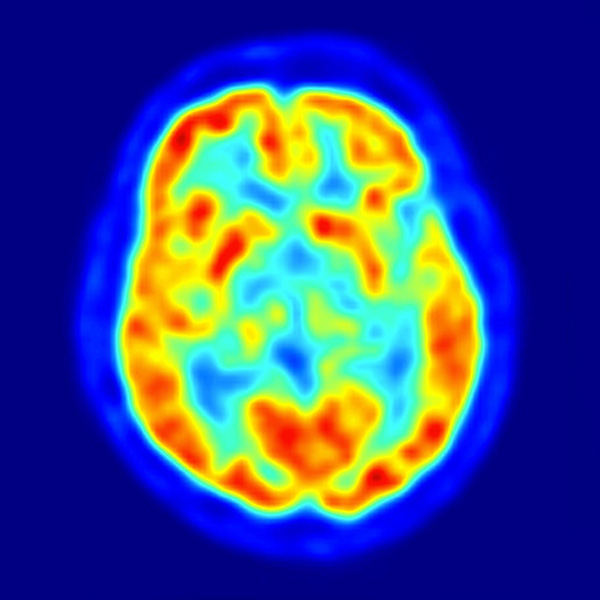
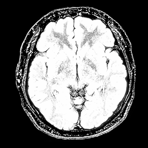
Clinicians today have to refer to a variety of medical images such as Magnetic Resonance Images (“MRI”), Computerized Tomography (“CT”) scans, Positron Emission Tomography (“PET”) scans, and many more, to have an insight of the patient’s internal organs. Each image provides a different set of data and clinicians are required to switch back-and-forth between different types of medical images and there are nuances that the clinician may miss in the process.
With Version 2.0 of the X³ 3D Physics Engine, BioMedX is merging the various different medical image data to form a comprehensive 3D model of the internal organ that can be applied to a variety of applications to optimize research, procedure, and treatment solutions.
Data Merge
Data merge is the fusion of various medical imaging data to form a comprehensive and personalized 3D model of a patient’s internal organ. Data merge provides clinicians an in-depth view through 3D models that can be used to diagnose, treat, and monitor patient conditions with the most precise and adaptable technology available.
Predictive Modeling
Version 2.0 of the X³ 3D Physics Engine provides predictive, personalized modeling as clinicians track and change critical data of the patient’s condition from initial diagnosis to the operating room to post- recovery monitoring. This delivers a more holistic approach to patient care and potentially leads to optimal procedural outcomes and speed of recovery.
Dynamic & Detailed
All medical imaging data are static. With a 3D digital model, additional variable datasets can be
introduced to make the model dynamic, enabling it to behave and respond accurately, which is critical in applications such as pre-surgical planning and other treatment simulations.Clinicians will be able to simulate a variety of conditions from surgery to treatment, therapy, and recovery.
Applications
Consultation Tool
As a consultation tool, clinicians can use a comprehensive 3D digital model to walk the patient through the procedure and address any concerns the patient might have. This includes showing the patient the 3D digital model of the internal organ and explaining the process details of the procedure. The clinician can rotate and annotate in a 3D environment to provide a more personal experience and enable a better consultation experience.
Pre-Surgical Planning Tool
For complex procedures, the comprehensive dynamic 3D digital model can be used as a pre- surgical planning tool for surgeons to rehearse the procedure prior to performing the actual surgery on the patient. This results in better preparation, while minimizing surgical time, risks, and costs, as well as optimizing the outcomes and reducing patient recovery time.
Intra-Operative Tool
In the future, as BioMedX advances the X³ 3D Physics Engine, there is the opportunity to develop intra-operative tools with it. By using Mixed Reality and/or Augmented Reality devices, the 3D digital organs can be projected directly onto the patient in the operating theatre. This enables surgeons to always have their “eyes on patient” and have all the critical medical imaging data in front of them. The tool can also serve as a roadmap to guide the surgeon through the surgery process to minimize the risks and optimize the outcomes.
Drug Research
With 3D digital models, pharmaceutical researchers can apply a variety of variable data to them to simulate the outcomes of various drug interactions. This provides a safe and effective way to test the efficacy and efficiency of the drug, and the simulations can be multiplied to mimic clinical trials to further validate the results.
Education
The 3D models can serve as digital cadavers for medical schools, enabling students to have repetitive access to multiple anatomical and physiological mock scenarios to expand their learning and training experiences.